High-resolution ultrasound from FUJIFILM VisualSonics allows you to visualize blood flow in an insect as small as a fruit fly (Drosophila) shown below. This video was captured using the MX700 probe on the Vevo 3100 ultrasound system in Color Doppler mode.
This recent study by Rutledge et al. compares 4D ultrasound (4D-US) with conventional M-mode and B-mode ultrasound for the assessment of LV function in a myocardial infarct model.
This study by Grune et al. compares LV functional analysis completed using a novel automated 2D-border detection algorithm (referred to as Auto2DE) versus conventional manual 2D echo assessment (2DE).
This study by Heinen et al. compares cardiac functional data in a myocardial infarct model acquired by B-Mode in parasternal long axis (PSLAX) versus Simpson method, versus MRI.
Researchers at the Mouse Imaging Centre of the Hospital for Sick Children in Toronto have established comprehensive methodologies for mouse cardiovascular imaging using high-frequency ultrasound successfully applying the established methodology to the phenotyping of mutant mouse models with human diseases.
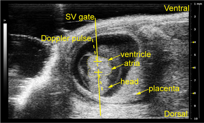
Using the Vevo 2100 and ultrasound pulsed-wave (PW) Doppler imaging, Caralynn Wilczewski from Dr. Frank L Conlon's lab has developed a reliable method for performing non-invasive in utero embryonic echocardiography on early gestation mouse embryos. Read more.